Last week we learned about vectors and I showed you the scale diagram method for solving a vector problem, such as determining the displacement of an object after a journey. The video is a short re-cap of the scale diagram technique.
By now you should have watched the video about satellites. This screenshot showing a satellite passing over the Highlands was taken from about 17 minutes into the programme – did you notice at the time?
It was quite eye-opening to see just how much modern society relies on satellite technology. I’ve got some more examples of the uses for satellites below.
A satellite moves horizontally at constant speed but also accelerates vertically towards the planet’s surface due to gravity. Thankfully, the curvature of the Earth means the satellite doesn’t crash but keeps on orbiting the planet.
Satellites can be used for environmental purposes, such as
In space there is no air resistance to oppose motion, so the Space Shuttle orbiter could travel at very high speeds, up to 17,000 mph! At these speeds, the orbiter experienced enormous air resistance as it descended into the Earth’s atmosphere at the end of its mission.
Air resistance is just like any other form of friction – it converts kinetic energy into heat energy. The effect of this heat energy is demonstrated in this video clip taken by a Canadian police car camera. It shows a meteor burning up in the atmosphere above Edmonton.
The high temperatures created during re-entry ionised the gas around the orbiter and this is often seen as a bright light in NASA cockpit videos, such as the one shown below.
To protect the vehicle and its crew from these high temperatures, the underside of the orbiter was covered by a layer of heat resistant tiles called thethermal protection system. This NASA clip explains how the tiles are constructed and arranged on the underside of the orbiter.
When Columbia was launched in 2003, something fell against the insulation on the left wing and knocked off some of the tiles. This hole in the thermal protection system caused Columbia to explode over the US as it re-entered the atmosphere. There is a wikipedia article about the Columbia disaster.
Video footage of NASA’s Houston control room from the morning of the disaster was included in the BBC Horizon documentary Final Descent – Last Flight of Space Shuttle Columbia.
WARNING: This last film is an excerpt from the Horizon programme and includes genuine cockpit video that was found in the wreckage, with some clips of the crew’s final minutes before they were killed.
There is a good description of the Space Shuttle at How Stuff Works.
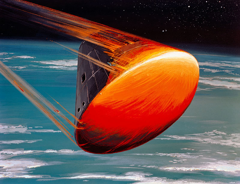
Before the space shuttle, each spacecraft was designed to be used only once and it was only the capsule containing the crew that returned to Earth. This was a small conical vehicle that had a thick heat shield on its base to withstand the heat of re-entry.
artist’s impression of Apollo capsule re-entering the atmosphere
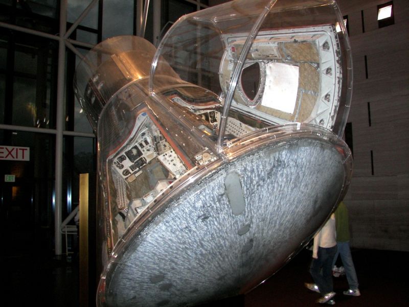
An ablative heat shield was used for these capsules. The material covering the base was designed to heat up until it sublimed (changed from solid to gas). The latent heat of sublimation is much greater than that required for fusion or vaporisation, so much more heat energy could be absorbed by the shield material as it changed state. Obviously there is a catch…the longer the shield protects the astronauts, the thinner it becomes! Here is an image of a Gemini IV capsule on display at the Smithsonian National Air and Space Museum showing what was left of the heat shield after successful return to Earth.
X-rays are a form of electromagnetic radiation. They have a much higher frequency than visible light or ultraviolet. The diagram below, taken from Wikipedia, shows where x-rays sit in the electromagnetic spectrum.
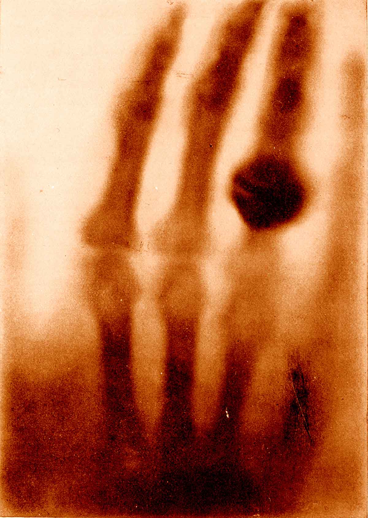
Wilhelm Röntgen discovered x-rays and the image below is the first x-ray image ever taken. It shows Mrs. Röntgen’s hand and wedding ring. The x-ray source used by Röntgen was quite weak, so his wife had to hold her hand still for about 15 minutes to expose the film. Can you imagine waiting that long nowadays?
This was the first time anyone had seen inside a human body without cutting it open. Poor Mrs. Röntgen was so alarmed by the sight of the image made by her husband that she cried out “I have seen my death!” Or, since she was in Germany, it might have been
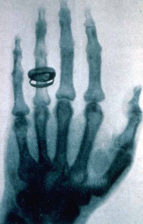
Röntgen continued to work on x-rays until he was able to produce better images. The x-ray below was taken about a year after the first x-ray and you can see the improvements in quality.
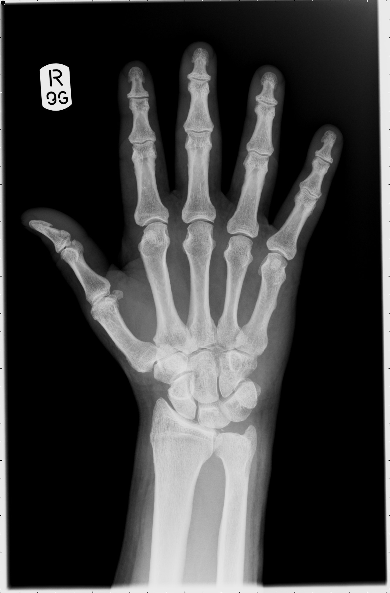
Notice that these early x-rays are the opposite of what we would expect to see today. They show dark bones on a lighter background while we are used to seeing white bones on a dark background, such as the x-ray shown below. The difference is due to the processing the film has received after being exposed to x-rays.
In hospitals, x-rays expose a film which is then developed and viewed with bright light. X-rays are able to travel through soft body tissue and the film behind receives a large exposure.
The x-rays darken the film.
More dense structures such as bone, metal fillings in teeth, artificial hip/knee joints, etc. block the path of x-rays and prevent them from reaching the film. Unexposed regions of the film remain light in colour.
Röntgen’s x-ray films would have involved additional processing steps. The exposed films were developed and used to create a positive. In creating a positive, light areas become dark and dark areas become light. So the light and dark areas in Röntgen’s x-rays are the opposite of what we see today. Our modern method makes it easier to detect issues in the bones as they are the lighter areas.
Röntgen was awarded the first ever Nobel Prize for Physics in 1901 for his pioneering work in this field of physics.
X-rays are a very high energy form of electromagnetic radiation. This means they have the potential to harm living cells. Medical staff only take x-rays of a patient when it is necessary to give a correct diagnosis but that wasn’t always the case.
I have attached a recording of a short BBC radio programme about the first x-ray and what people in the Victorian era thought of these new images. Click on the player at the end of this post.
This week, we’ve looked at calculating radiation doses. The absorbed dose D, measured in Grays (Gy), takes into account the energy E absorbed and the mass m of the absorbing tissue.
The higher the energy, the greater the absorbed dose. If you are wondering why the absorbing mass is important, consider the different masses of tissue involved in a dental x-ray and a chest x-ray….
We also learned about equivalent dose in Sieverts (Sv). The equivalent dose H gives an indication of the potential for biological harm by considering the absorbed dose D and a weighting factor .
Different types of radiation have different weighting factors, e.g.
type of radiation
weighting factor
gamma
1
x-ray
1
beta
1
alpha
20
The more damaging forms of radiation have a larger weighting factor.
Absorbed dose and equivalent dose are usually expressed in smaller units; μGy, mGy, μSv, mSv.
In the UK, the Government has set an effective equivalent dose of 1mSv per year for members of the public. This limit can be increased to 20mSv for people who work in the nuclear industry, certain medical occupations (such as radiographers) and airline pilots – all of whom will exceed the public limit in the course of their job.
This occupational increase for some individuals can be justified on the grounds that workers are not as vulnerable to the effects of radiation exposure since they are neither children (high rate of cell division so more chance of dna damage being copied) or elderly (reduced ability to repair damage). In many cases, these workers will also be screened on a regular basis by occupational health staff at their place of work.
Here is a poster from the excellent xkcd site that explores examples of the different levels of equivalent dose.
We started looking at half-life today. The attached file walks you through different types of half-life problem. There are some questions for you to try along they way. The answers are at the end, please don’t cheat!
The Geiger-Müller (GM) counter is used to detect ionising radiation such as alpha and beta particles or gamma rays. The radiation enters through a very thin window at one end of the tube. This window is usually made of mica.
Mica is a mineral that forms in layers called sheets. These sheets can be split apart into very thin layers, so thin that even an alpha particle can pass through it (remember that alpha particles can be stopped by something as thin as your skin or a sheet of paper). The mica window prevents the argon inside the tube from escaping and also stops air from getting into the tube.
When radiation enters the tube and collides with an argon atom, an electron may be knocked off the atom – we call this process ionisation. When ionisation occurs, a positively-charged argon ion and a negatively-charged electron are produced. The argon ion is attracted to the outside wall of the tube, which is connected to the negative terminal of the power supply, while the electron is attracted to the central electrode, which is kept at a high positive voltage – typically 500V.
A small pulse of current is produced each time an electron reaches the central electrode. These pulses can be counted by an electronic circuit and a displayed on a 7-segment display. Sometimes a small speaker is added to the system to produce a click for each pulse. On its own, the GM tube cannot tell the difference between alpha, beta and gamma radiation. We need to place different materials (e.g. paper, aluminium, lead) in front of the mica window to discover which type of radiation is responsible for the reading.
Here is a short video demonstrating the use of a Geiger-Müller tube.
X-rays are a form of electromagnetic radiation. They have a much higher frequency than visible light or ultraviolet. The diagram below, taken from Wikipedia, shows where x-rays sit in the electromagnetic spectrum.
Wilhelm Röntgen discovered x-rays and the image below is the first x-ray image ever taken. It shows Mrs. Röntgen’s hand and wedding ring. The x-ray source used by Röntgen was quite weak, so his wife had to hold her hand still for about 15 minutes to expose the film. Can you imagine waiting that long nowadays?
This was the first time anyone had seen inside a human body without cutting it open. Poor Mrs. Röntgen was so alarmed by the sight of the image made by her husband that she cried out “I have seen my death!” Or, since she was in Germany, it might have been
Röntgen continued to work on x-rays until he was able to produce better images. The x-ray below was taken about a year after the first x-ray and you can see the improvements in quality.
Notice that these early x-rays are the opposite of what we would expect to see today. They show dark bones on a lighter background while we are used to seeing white bones on a dark background, such as the x-ray shown below. The difference is due to the processing the film has received after being exposed to x-rays.
In hospitals, x-rays expose a film which is then developed and viewed with bright light. X-rays are able to travel through soft body tissue and the film behind receives a large exposure. The x-rays darken the film. More dense structures such as bone, metal fillings in teeth, artificial hip/knee joints, etc. block the path of x-rays and prevent them from reaching the film. Unexposed regions of the film remain light in colour.
Röntgen’s x-ray films would have involved additional processing steps. The exposed films were developed and used to create a positive. In creating a positive, light areas become dark and dark areas become light. So the light and dark areas in Röntgen’s x-rays are the opposite of what we see today. Our modern method makes it easier to detect issues in the bones as they are the lighter areas.
Röntgen was awarded the first ever Nobel Prize for Physics in 1901 for his pioneering work in this field of physics.
I have attached a recording of a short BBC radio programme about the first x-ray and what people in the Victorian era thought of these new images. Click on the player at the end of this post or listen to it in iTunes.


 .
.


