The attached pdf document contains notes to help with your revision of the national 5 waves and radiation unit. Your unit assessment is scheduled for next week (see calendar dates on right on screen). Additional revision materials are available on BBC Bitesize.
 mrmackenzie

Higher unit 3 solutions to practice NAB
Here are the solutions to the practice NAB. Please try the questions before looking at this file. Any questions that can’t wait ’til the start of term? Try the BBC Bitesize Radiation & Matter site or my Higher page.
Higher unit 3 practice NAB
Here is the practice NAB for unit 3. Please download these questions and use them to gauge your readiness for the nab, which will be in the first week of term – see dates under the calendar.
national 4 electricity & energy solutions
Here are the solutions to the electricity and energy questions.
Remember that these sample questions & answers do not cover all of the key areas within the unit and you should work through the notes I provided earlier.
national 4 electricity & energy KU revision
Here are some old exam questions to help you to prepare for the resit of the E&E unit assessment. Your resit is scheduled for Thursday of this week (see calendar dates on right of the screen). You will get more thorough coverage of the appropriate key areas if you work through the BBC Bitesize pages and try their tests.
Download the attached questions if you feel ready to try making accurate statements. I will post my answers on Monday evening.
national 4 dynamics & space solutions
Here are the solutions to the revision questions. Please try answering the questions before downloading these answers.
national 4 dynamics & space KU revision
If you have to resit any of the national 4 unit assessments, the test paper will only contain knowledge questions. The revision material on BBC Bitesize has multiple choice test questions but you should also practise making accurate statements if you want to pass the test.
Here is a selection of suitable questions I have extracted from old SQA papers. Work through them and compare your responses to those given in the answer booklet (see next post).
Your Dynamics and Space resit will take place on Monday 30th March.
equivalent dose
This week, we’ve looked at calculating radiation doses. The absorbed dose, measured in Grays, takes into account the mass of the absorbing tissue, while the equivalent dose (in Sieverts) gives an indication of the potential for biological harm.
Equivalent dose is calculated using a weighting factor. More damaging forms of radiation have a larger weighting factor.
Absorbed dose and equivalent dose are usually expressed in smaller units; uGy, mGy, uSv, mSv.
Here is a poster from the excellent xkcd site that explores examples of the different levels of equivalent dose.
Click on the picture for a larger version.

Notice that the scale changes as you move through the poster from blue to green to red.
The dosimetry topic is very short and comprehensively covered at BBC Bitesize.
viewing the solar eclipse
We spoke about the eclipse in class today. It’s really important that you follow advice for watching the eclipse safely to avoid permanent damage to your eyes.
You can buy eclipse viewing glasses online. They look like this
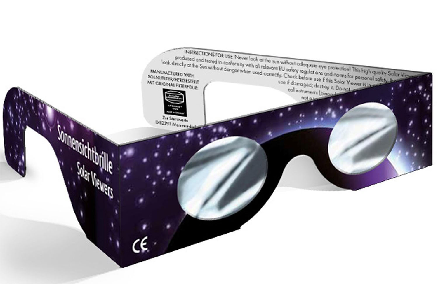
and only cost a few pounds per pair. Make sure any glasses you buy have the CE mark on them.
Here is one UK retailer of eclipse viewing glasses who still has them in stock.
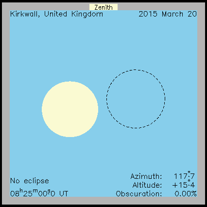
Although we’re not in the totality (100%) zone, Thurso is well placed to get a good view. Here is a simulation for Orkney, our view will be very similar. Click on the picture to start to animation.
x-rays
X-rays are a form of electromagnetic radiation. They have a much higher frequency than visible light or ultraviolet. The diagram below, taken from Wikipedia, shows where x-rays fit into the electromagnetic spectrum.

image by Materialscientist
Wilhelm Röntgen discovered x-rays and the image below is the first x-ray image ever taken. It shows Mrs. Röntgen’s hand and wedding ring. The x-ray source used by Röntgen was quite weak, so his wife had to hold her hand still for about 15 minutes to expose the film. Can you imagine waiting that long nowadays?

This was the first time anyone had seen inside a human body without cutting it open. Poor Mrs. Röntgen was so alarmed by the sight of the image made by her husband that she cried out “I have seen my death!” Or, since she was in Germany, it might have been
“Ich habe meinen Tod gesehen!“
that she actually said.
Röntgen continued to work on x-rays until he was able to produce better images. The x-ray below was taken about a year after the first x-ray and you can see the improvements in quality.

Notice that these early x-rays are the opposite of what we would expect to see today. They show dark bones on a lighter background while we are used to seeing white bones on a dark background, such as the x-ray shown below. The difference is due to the processing the film has received after being exposed to x-rays.
 In hospitals, x-rays expose a film which is then developed and viewed with bright light. X-rays are able to travel through soft body tissue and the film behind receives a large exposure. The x-rays darken the film. More dense structures such as bone, metal fillings in teeth, artificial hip/knee joints, etc. block the path of x-rays and prevent them from reaching the film. Unexposed regions of the film remain light in colour.
Röntgen’s x-ray films would have involved additional processing steps. The exposed films were developed and used to create a positive. In creating a positive, light areas become dark and dark areas become light. So the light and dark areas in Röntgen’s x-rays are the opposite of what we see today. Our modern method makes it easier to detect issues in the bones as they are the lighter areas.
Röntgen was awarded the first ever Nobel Prize for Physics in 1901 for his pioneering work in this field of physics.
Medical imaging has come a long way since Röntgen’s discovery of x-rays. This promotional video from German company Siemens outlines the advances that have been made since the early 20th century.
I have attached a recording of a short BBC radio programme about the first x-ray and what people in the Victorian era thought of these new images. Click on the player at the end of this post or listen to it in iTunes.
